Connecting Brains
YouTube search... ...Google search
- Symbiotic Intelligence ... Connecting Brains ... Neuroscience ... Nanobots
- Biocomputing ... Bio-inspired Computing ... Neuromorphic ... Molecular ... (Artificial) Immune System ... Self-Organizing
- Three-Second Pause short story
- Life~Meaning ... Consciousness ... Creating Consciousness ... Biology ... Quantum Biology ... Proteins ... LEV ... TAME ... Orch-OR
- Life Sciences ... Evolutionary ... Bioinformatics ... Proteins ... Genome ... Genealogy ... Pharmaceuticals ... Chemistry
- Apprenticeship Learning - Inverse Reinforcement Learning (IRL)
- Healthcare ...Brain and Spine
- Artificial Intelligence (AI) ... Generative AI ... Machine Learning (ML) ... Deep Learning ... Neural Network ... Reinforcement ... Learning Techniques
- Collective Animal Intelligence ... Animal Ecology ... Animal Language ... Bird Identification
- Gaming ... Game-Based Learning (GBL) ... Security ... Generative AI ... Games - Metaverse ... Quantum ... Game Theory ... Design
- Neuroscience News - Bain Computer Interface
- Artificial intelligence turns brain activity into speech | Kelly Servick
- 29 Neurotech Companies Interfacing With Your Brain | Nanalyze
- CTRL-labs Uses AI for Brain-Computer Interface | Nanalyze
- Synchron – A Brain Computer Interface Using Stentrodes | Nanalyze
- Researchers translate brain waves into scarily accurate images using Stable Diffusion AI | Katie Wickens - PC Gamer ... Researchers have been able to use Stable Diffusion models to reconstruct images from human brain waves.
- Scientists recorded a Pink Floyd song from patients’ brain waves. The tech could eventually allow for communication without words | Erin Prater - Fortune Well ... While receiving surgery they hoped would cure intractable seizures, Pink Floyd’s 1979 single “Another Brick in the Wall, Part 1” played in the operating room. Using artificial intelligence, Bellier was able to reconstruct the song from that electrical activity in each patient’s brain
- ‘Biocomputer’ combines lab-grown brain tissue with electronic hardware | Lilly Tozer - Nature ... A system that integrates brain cells into a hybrid machine can recognize voices.
- Forget Neuralink: The Real Brain Tech Trade Has 10 Names You’ve Never Heard | Jonathan Rose
Connecting brains encompasses technologies that measure neural activity, translate it into useful outputs, deliver information back to the nervous system, or model the circuits that make these processes possible. A brain-computer interface (BCI), also called a brain-machine interface (BMI), establishes a functional pathway between brain activity and an external device. A brain-to-brain interface adds a second person's nervous system to that pathway, while connectomics maps neural wiring and whole brain emulation investigates whether computational models can reproduce brain functions. These fields connect Neuroscience, machine learning, healthcare, and bio-inspired computing, but they address different scientific problems.
The clearest demonstrated benefits concern restoring communication and control to people with severe motor impairments. Research participants have used neural interfaces to operate computers, generate text or synthesized speech, control robotic devices, and interact with stimulation systems that assist movement. Those achievements do not establish unrestricted access to private thoughts, downloadable skills, or the transfer of a person's consciousness into a computer. The evidence summarized here includes peer-reviewed studies and explicitly identified company reports available through October 27, 2026.
Whole Brain Emulation and the Connectome
A connectome describes connections among neurons or brain regions at a specified scale. At synaptic resolution, researchers use electron microscopy, automated image segmentation, and extensive proofreading to reconstruct neurons and identify their contacts. The result can be represented as a graph, with neurons as nodes and connections as edges, but an anatomical connection does not completely specify its physiological effect. Synaptic strength, neurotransmitters, cellular properties, and the organism's current state influence what the circuit actually does.
In 2024, the FlyWire collaboration published a wiring diagram of an adult female Drosophila melanogaster brain containing 139,255 reconstructed neurons and approximately 50 million chemical synapses between them. This was a major advance beyond earlier partial adult-fly reconstructions and the much smaller larval brain connectome. The brain dataset should not be confused with a complete account of the animal's peripheral nervous system, muscles, or ventral nerve cord. Its greatest immediate value is enabling researchers to trace circuits, compare cell types, and formulate experimentally testable explanations of behavior.
- Neuronal wiring diagram of an adult brain | Dorkenwald and colleagues, Nature, 2024 Primary description of the FlyWire adult-fly brain reconstruction.
- Whole-brain annotation and multi-connectome cell typing of Drosophila | Schlegel and colleagues, Nature, 2024 Cell classifications and comparisons that make the wiring diagram biologically interpretable.
By 2026, connectomics had expanded beyond the isolated adult brain to datasets spanning the brain and ventral nerve cord. The BANC study examined distributed control circuits across an adult female fly's brain-and-cord connectome, while a male central-nervous-system reconstruction enabled complementary comparisons. These resources help investigate how descending commands interact with local motor circuits and how anatomy varies across individuals and sexes. They also make clear that the anatomical scope of a particular dataset must be stated rather than inferred from the phrase “whole brain.”
- Distributed control circuits across a brain-and-cord connectome | Bates, Phelps, Kim, Yang, and colleagues, Nature, 2026 Connectomic analysis spanning brain and ventral nerve cord.
- World-first map of a male fly brain a win for neuroscience | UK Research and Innovation, September 4, 2026 Institutional account of the male central-nervous-system reconstruction and comparative research.
Emulated Behavior and Technical Limitations
Whole brain emulation aims to reproduce relevant brain functions in a computational system, but researchers disagree about the necessary level of biological detail and the tests that would establish success. A simulation may reproduce selected responses without preserving an animal's full repertoire, developmental history, or memories. Consequently, a convincing animation is insufficient evidence of a faithful neural replica. Stronger evaluation requires predictions of neural activity and behavior that survive comparison with experiments, including interventions not used to construct the model.
In March 2026, Eon Systems demonstrated a virtual fly combining a connectome-constrained neural model with the NeuroMechFly body simulation. Eon's technical account describes simplified spiking neurons, a small set of descending control signals, and existing movement controllers trained to imitate fly behavior. Several brain-to-body mappings were chosen by hand, while learning, plasticity, and changing internal biological states were largely absent. The demonstration is therefore a useful example of embodied modeling and imitation-based control, but it does not show that wiring alone recreates an individual animal or transfers consciousness.
- The First Multi-Behavior Brain Upload | Eon Systems, March 7, 2026 Company announcement of the embodied-fly demonstration; its terminology expresses the company's interpretation.
- How the Eon Team Produced a Virtual Embodied Fly | Eon Systems, March 10, 2026 Technical explanation of the component models, controller dependence, hand-selected mappings, and unresolved limitations.
Historical commentary: The First Brain Was Just Uploaded To A Computer
The title of this retained video uses “uploaded” more broadly than a demonstrated transfer of a living mind. Read it alongside the technical limitations above and the underlying research rather than treating its title as an established scientific conclusion. Behavioral resemblance, circuit reconstruction, and personal continuity are separate claims requiring different evidence.
Philosophical and Ethical Challenges
Human emulation raises questions about personal identity that technical performance alone cannot settle. Even a hypothetical model with convincing memories and behavior would leave open whether it continues the original person's experience or constitutes a new individual. Current serial-section electron-microscopy workflows require fixed tissue and destructive preparation, but this does not prove that every conceivable future brain-measurement method must destroy its subject. No validated procedure currently demonstrates the transfer of a human person's identity or consciousness into software.
Questions of digital welfare also require care because neither realistic behavior nor an elaborate neural model establishes the capacity to experience suffering. Ethical analysis can consider precautionary safeguards without assuming that present simulations are sentient. Relevant concerns include who could create, modify, copy, suspend, or delete a hypothetical emulation and how consent could remain meaningful under those conditions. These issues connect to ethics, transhumanism, and personal identity and meaning, while remaining distinct from the immediate clinical responsibilities of BCI research.
Brain Machine Interface (BMI) / Brain Computer Interface (BCI)
YouTube search... ...Google search
- Kernel Optical neuroimaging technology and services; measurement capabilities should be distinguished from demonstrated clinical communication systems.
- Brain-Computer Interfaces (BCI) | Singularity Hub Historical and continuing reporting on neural interfaces.
- Brain-computer interface research using inverse reinforcement learning | Tanushree Shenwai, MarkTechPost, 2022 Historical coverage connecting BCI control to inverse reinforcement learning.
Recording Methods and the Signal Path
A BCI measures a limited sample of brain activity and learns how that sample relates to a task. Recording hardware captures the signal, signal processing reduces interference, and a decoder estimates an intended command or communication output. The user receives visual, auditory, or tactile feedback and adjusts subsequent activity, forming a feedback loop. Both the person and the software may learn, so useful performance depends on their interaction rather than the decoder alone.
| Method | What it measures | Advantages | Important constraints |
|---|---|---|---|
| Electroencephalography (EEG) | Electrical potentials measured at the scalp | Noninvasive, portable, and temporally precise | Signals mix across sources and are vulnerable to eye, muscle, movement, and environmental interference. |
| Electrocorticography (ECoG) | Electrical activity recorded on or near the cortical surface | Stronger local signals than scalp EEG and useful coverage of cortical populations | Implantation requires an invasive procedure; performance depends on placement and long-term stability. |
| Intracortical microelectrodes | Spiking activity and other local electrical signals within cortex | Detailed information useful for movement and speech decoding | Surgery, tissue response, electrode movement, and hardware longevity remain central concerns. |
| Endovascular electrodes | Electrical activity recorded from electrodes positioned in a blood vessel near cortex | Access through the vascular system can avoid direct insertion of electrodes into brain tissue | It remains an invasive implant procedure; vessel anatomy, clotting risks, and signal access constrain deployment. |
| Functional MRI (fMRI) | Blood-oxygenation changes associated with neural activity | Broad spatial coverage without a brain implant | Indirect, delayed signals and a large scanner limit portability and conversational responsiveness. |
| Functional near-infrared spectroscopy (fNIRS) | Optical changes related to cortical blood oxygenation | Potentially wearable measurement without surgery | Limited depth, systemic physiological interference, and delayed hemodynamic responses require careful interpretation. |
Noninvasive does not mean that a system reads every thought or functions without calibration. EEG interfaces often use motor imagery, attention to flickering targets, or responses to selected stimuli, each with different demands on the user. Hemodynamic methods measure consequences of neural activity rather than individual spikes, so their information and timing differ from implanted electrical recordings. A demonstration with one modality cannot establish that another modality will support the same vocabulary, speed, or accuracy.
Surface electromyography (sEMG) is an important neighboring technology but records muscle activity rather than brain activity. Work associated with CTRL-labs and Meta has developed wrist-based interfaces that infer intended actions from these peripheral signals, including a substantial 2025 study. Such systems can provide useful wearable input without brain surgery, but their success should not be described as direct cortical decoding. The distinction matters particularly for users whose disease or injury interrupts signals before they reach the muscles.
- A generic non-invasive neuromotor interface for human-computer interaction | Nature, 2025 Primary research on computer input decoded from surface electromyography.
Decoding, Calibration, and Reliable Evaluation
Decoders range from statistical classifiers and regression models to recurrent networks and other deep learning architectures. Training associates recorded activity with known targets, such as an attempted movement, phoneme, or selected symbol. Preprocessing, accurate timing, and appropriate training labels can be as important as model size. Online adaptation may help maintain performance, but it must avoid reinforcing errors when the user's intended output is uncertain.
Neural recordings vary across people and sessions because of physiology, attention, fatigue, recording changes, and the task itself. A model that succeeds on randomly mixed samples may fail on a genuinely later session, making temporal separation of training and testing essential. Evaluation should report the participant count, vocabulary or action space, calibration time, error corrections, latency, and whether testing was offline or live. Useful comparisons also include existing assistive technologies and the effort required from the user and caregivers.
Headline accuracy figures can hide very different achievements. Classifying one of two choices is easier than producing unrestricted sentences, and a language model may improve plausible wording without faithfully preserving the intended message. Words per minute, word error rate, task completion, false activations, and reliable hours of independent use answer different questions. This makes verification and validation and protection against overfitting central to assessing whether a laboratory demonstration can become a dependable assistive system.
Restoring Movement and Sensation
Motor BCIs can translate attempted movement into cursor motion, robotic-arm control, or commands to an external stimulator. Functional electrical stimulation can activate muscles through electrodes, while spinal stimulation engages circuits below an injury. These approaches bypass or assist impaired pathways rather than necessarily repairing the original damage. The NeuroLife work at Battelle and Ohio State is a historical example of connecting cortical recordings to muscle stimulation, while later brain-spine systems extend the principle to coordinated movement.
In a 2023 Nature study, a brain-spine interface linked cortical recordings to epidural spinal stimulation in one participant with chronic spinal cord injury. The system supported standing and walking through a combination of implanted technology, decoding, stimulation, and rehabilitation. This was a major proof of concept rather than evidence that every person with paralysis can recover the same function. Bidirectional interfaces also investigate stimulation that supplies useful sensory feedback, which could help users adjust grip and movement without relying entirely on vision.
- Walking naturally after spinal cord injury using a brain-spine interface | Lorach and colleagues, Nature, 2023 Human proof of concept linking cortical signals to spinal stimulation.
- A brain-computer interface that evokes tactile sensations improves robotic arm control | Flesher and colleagues, Science, 2021 Primary human study demonstrating the benefit of stimulation-mediated tactile feedback during robotic manipulation.

Recent Developments
Speech restoration has progressed from constrained demonstrations toward larger vocabularies and more responsive communication. A 2024 UC Davis study in a participant with ALS reported approximately 97.5% correct word decoding after additional calibration, using attempted speech rather than unrestricted thought extraction. In 2025, a Berkeley/UCSF system generated streaming speech in short chunks instead of waiting for an entire sentence. A separate UC Davis study demonstrated real-time voice synthesis with controllable intonation, although listener intelligibility remained substantially below perfect speech.
These results should be interpreted through their individual study designs rather than combined into a single performance claim. Text accuracy, audio intelligibility, speech rate, and expressive control measure different parts of communication. Small participant samples and specialized support also limit conclusions about broad clinical availability. Nevertheless, the progression toward continuous output and recognizable personal voices is meaningful for speech synthesis, autonomy, and everyday interaction.
- Brain-computer interface helps paralyzed man speak | NIH, September 10, 2024 Summary of the UC Davis study, including calibration and approximately 97.5% word accuracy.
- A streaming brain-to-voice neuroprosthesis to restore naturalistic communication | Nature Neuroscience, 2025 Primary study of low-latency streaming synthesis from cortical recordings.
- First-of-its-kind technology helps man with ALS speak in real time | UC Davis Health, June 11, 2025 Explains expressive voice synthesis and reports that listeners understood almost 60% of synthesized words in the reported evaluation.
Historical BCI Lectures and Demonstrations
The following retained videos document earlier explanations, experiments, and expectations. Their publication-era descriptions do not establish current clinical availability, and predictions of general enhancement should be distinguished from the specific capabilities discussed above. Historical references to employers, academic positions, and future products should be read in their original context.
|
|
|
|
|
|
|
|
|
|
|
|
|
|
|
|
|
|
Brain Machine Interface (BMI) via Generative AI
Generative AI can help convert noisy neural measurements into fluent language, plausible images, or recognizable sound. Such systems combine evidence from a participant's recordings with patterns learned from other data, including the statistical structure of language or images. This is useful because the recorded signal is incomplete, but it also means that an output may contain information supplied by the model rather than directly recovered from the brain. Generated detail must therefore be distinguished from measured evidence, especially when evaluating apparent “mind reading.”
In 2020, Makin, Moses, and Chang demonstrated an encoder-decoder approach to translating cortical recordings during overt speech into text under constrained experimental conditions. This connected neural decoding to encoder-decoder methods and sequence modeling. It did not establish a general device that transcribes arbitrary silent thoughts. Later communication systems build on related ideas while changing the recording method, training data, vocabulary, and intended user population.
- Machine translation of cortical activity to text with an encoder-decoder framework | Makin, Moses, and Chang, Nature Neuroscience, 2020 Foundational study requiring careful distinction between constrained overt-speech decoding and unrestricted thought transcription.
Semantic, Visual, and Musical Reconstruction
Tang and colleagues' 2023 semantic decoder used fMRI and a language model to reconstruct aspects of the meaning of perceived or imagined language. The principal experiments required extensive participant-specific training, approximately 16 hours of listening data, and generally recovered meaning more reliably than exact wording. Participants could disrupt decoding through competing mental activities, and models did not simply transfer to unfamiliar individuals. The result demonstrated an important link between language modeling and brain measurements, not a portable, covert, universal thought reader.
Visual reconstruction similarly combines a measured neural signal with a powerful learned prior. Takagi and Nishimoto's 2023 work used latent diffusion models to reconstruct images from fMRI responses to viewed pictures. A visually convincing reconstruction can include plausible features that were not accurately recovered, making independent stimulus tests and quantitative comparisons necessary. These studies concern controlled visual experiences and should not be generalized to reliable recordings of arbitrary dreams or private memories.
Music reconstruction provides a complementary example involving auditory cortex. Bellier and colleagues reconstructed a recognizable version of a Pink Floyd recording from intracranial electrical activity obtained while patients listened to it. The experiment used electrodes placed for clinical monitoring, not a consumer EEG headset, and the reconstructed audio was imperfect. Its significance concerns neural representation of music and nonlinear decoding, rather than proof that an arbitrary remembered song can be extracted on demand.
- Semantic reconstruction of continuous language from non-invasive brain recordings | Tang, LeBel, Jain, and Huth, Nature Neuroscience, 2023 Primary semantic-decoding study.
- Brain Activity Decoder Can Reveal Stories in People's Minds | UT Austin, May 1, 2023 Institutional explanation of training requirements, cooperation, and limitations.
- High-Resolution Image Reconstruction With Latent Diffusion Models From Human Brain Activity | Takagi and Nishimoto, CVPR 2023 Primary paper on diffusion-assisted reconstruction from fMRI.
- Music can be reconstructed from human auditory cortex activity using nonlinear decoding models | Bellier and colleagues, PLOS Biology, 2023 Primary intracranial music-reconstruction study.
Inner Speech and Control Over Output
A 2025 study extended speech decoding to imagined inner speech in four participants with severe speech and motor impairments. Inner speech produced distinguishable, generally weaker activity than attempted speech, and decoding remained imperfect. The researchers also demonstrated approaches to reject unintended inner speech and to activate an inner-speech decoder using an imagined password. These are useful safeguards under the tested conditions, not evidence that all future mental-privacy risks are solved.
For communication systems, a fluent sentence must remain subordinate to the user's intent. A decoder can misidentify sounds, and a language model can turn uncertain evidence into an apparently confident statement. Interfaces therefore need ways to pause output, correct errors, distinguish assistance from direct decoding, and obtain confirmation for consequential actions. This is a concrete role for human oversight, rather than a reason to assume that machine-generated wording is an authentic expression of its user's beliefs.
- Decoding inner speech from brain signals | NIH, September 9, 2025 Study summary reporting participant count, vocabulary-dependent error rates, and privacy safeguards.
- Scientists develop interface that reads thoughts from speech-impaired patients | Stanford, August 14, 2025 Researcher explanation distinguishing attempted speech, inner speech, and prevention of unintended output.
|
|
Neuralink
YouTube search... ...Google search
- Neuralink Homepage
- Neuralink | Wikipedia
- Neuralink Progress Update: What To Know About Elon Musk's Major Event | Inverse Historical reporting associated with earlier company presentations.
Neuralink develops implanted neural interfaces and robotic insertion technology. Its early public presentations included recordings from a pig and descriptions of thin electrode threads, while later human research focused on enabling people with severe paralysis to control computers. Those stages should be distinguished: recording neural signals in an animal is not the same as establishing durable human benefit. Likewise, a company objective involving vision, generalized enhancement, or human-AI integration is not itself a demonstrated clinical outcome.
The company's PRIME study describes an investigational implant and application for computer control. In January 2026, Neuralink announced 21 participants across its programs, and University College London Hospitals separately reported seven participants in GB-PRIME. These dated reports establish expansion of research participation rather than universal safety, effectiveness, or general commercial availability. Independent follow-up, transparent adverse-event reporting, and meaningful outcomes remain necessary when comparing implant systems.
In an October 1, 2026 update, Neuralink reported pretraining on 50,000 hours of unlabeled brain data and described improved decoder stability. This is relevant to self-supervised learning and the burden of repeated calibration, but the public update is a company report. Its performance claims should be evaluated against the task, participants, comparator, and follow-up period rather than treated as universal BCI benchmarks. Clinical usefulness ultimately depends on sustained benefit and manageable support needs as well as decoding performance.
- PRIME Study brochure | Neuralink Investigational study purpose and device description.
- Two Years of Telepathy | Neuralink, January 28, 2026 Company report announcing 21 participants.
- Seven GB-PRIME patients now participating in Neuralink trial | UCLH, January 29, 2026 Hospital report on the British trial.
- Pretraining on 50,000 Hours of Unlabeled Brain Data | Neuralink, October 1, 2026 Company account of self-supervised decoder development.
Other Implant Approaches
Neuralink is one part of a broader research landscape that includes academic BrainGate collaborations, cortical-surface recording systems, and endovascular devices. Synchron's Stentrode records through electrodes deployed in a cerebral blood vessel rather than threads inserted into cortex. Its published SWITCH study assessed an implanted system in four participants with severe paralysis, illustrating both the feasibility of this approach and the small scale of early evidence. Differences in surgical access, signal quality, supported tasks, and maintenance make simple “winner” rankings scientifically misleading.
- Assessment of Safety of a Fully Implanted Endovascular Brain-Computer Interface for Severe Paralysis in 4 Patients | Mitchell and colleagues, JAMA Neurology, 2023 The SWITCH study provides early human safety evidence for the endovascular approach.
Historical Neuralink Presentations
These retained presentations document earlier device concepts, animal demonstrations, and public expectations. Predictions about implantation dates or inevitable human enhancement should be read as statements made at the time, not current technical specifications. The dated research sources above provide the appropriate context for assessing subsequent progress.
|
|
|
|
|
|
Brain to Brain Interface (BBI)
YouTube search... ...Google search
- Scientists Demonstrate Direct Brain-to-Brain Communication in Humans | Robert Martone, Scientific American Historical reporting on experiments sometimes described as an “Internet of brains.”
A brain-to-brain interface combines neural recording, computer processing, a communication channel, and stimulation or another input to a second nervous system. Human demonstrations have often used EEG to select a binary signal and transcranial magnetic stimulation to produce a percept such as a flash of light. Participants interpret that percept through an agreed code, so the system transmits a limited message rather than directly sharing a complete thought. The intervening hardware and learned interpretation are essential parts of the communication pathway.
BrainNet, published in 2019, linked groups of three people performing a simplified collaborative game. Two participants supplied decisions through EEG, and a third received stimulation-mediated information to help choose an action. The study demonstrated constrained collaboration with a low-bandwidth channel, not the sharing of memories, language, or consciousness. Earlier animal experiments and human proof-of-concept studies remain useful historical steps, but their tasks, recording methods, and species must not be conflated.
The central technical challenges include reliable encoding, safe stimulation, feedback, and distinguishing neural information transfer from ordinary sensory cues. Ethical questions concern the recipient's ability to understand, authorize, and interrupt incoming stimulation. A communication system that affects another person's nervous system requires explicit boundaries around control, responsibility, and data handling. These issues connect BBI research to cybersecurity and privacy, even when the transmitted messages are only simple decisions.
- BrainNet: A Multi-Person Brain-to-Brain Interface for Direct Collaboration Between Brains | Jiang and colleagues, Scientific Reports, 2019 Primary human experiment combining EEG, computer communication, and brain stimulation.
|
|
|
|
|
|
PEDOT and Neural Interface Materials
- PEDOT:PSS Interfaces Support the Development of Neuronal Synaptic Networks with Reduced Neuroglia Response In vitro | Cellot and colleagues Laboratory evidence about neuronal cultures and glial responses, not proof of scar-free human implants.
- Chronic Intracortical Neural Recordings Using Microelectrode Arrays Coated With PEDOT-TFB | NeuroNexus
- Improving the performance of poly(3,4-ethylenedioxythiophene) for brain-machine interface applications | Mandal and colleagues, Acta Biomaterialia, 2014
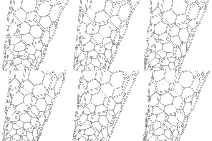
- Carbon Nanotube (CNT) | Wikipedia Background on cylindrical carbon structures considered in some electrode-material research.
An electrode must couple electronic hardware to tissue whose signals depend on ions moving through fluid and cell membranes. Its performance depends on surface chemistry, electrical impedance, charge transfer, mechanical properties, and the tissue response to implantation. Small recording sites improve spatial selectivity but can be difficult to connect to low-noise electronics, while stiff structures may move differently from surrounding tissue. Materials research therefore complements software advances rather than merely providing a passive support for the decoder.
PEDOT, poly(3,4-ethylenedioxythiophene), is a conducting polymer investigated for neural electrodes and other bioelectronic interfaces. In PEDOT:PSS, positively charged PEDOT is associated with negatively charged poly(styrenesulfonate), helping form a processable conducting material. Appropriate formulations and coatings can increase effective surface area and improve electrode-interface properties. Their behavior depends on fabrication, the supporting electrode, the biological environment, and the intended recording or stimulation task.
Promising cell-culture results do not establish that a coating eliminates scarring in a chronically implanted human device. Researchers must also evaluate adhesion, cracking or delamination, sterilization, electrochemical stability, degradation products, and inflammatory responses over time. Biofunctionalization with peptides or other molecules offers ways to modify interactions with tissue, but each formulation requires its own evidence. Earlier claims that one polymer would enable unrestricted brain-AI merging or full-bandwidth brain streaming went well beyond the cited material experiments.

Mapping the Brain
Brain mapping combines complementary measurements rather than producing a single all-purpose picture. Structural imaging describes anatomy, functional imaging measures activity-related changes, cell atlases distinguish molecularly defined populations, and connectomics traces physical connections. Linking these scales requires compatible data representations, spatial registration, and careful accounting for differences among specimens. A gene-expression map, an MRI atlas, and a synapse-level reconstruction answer different questions even when all are called a “brain atlas.”
Major resources include the NIH BRAIN Initiative, the BRAIN Initiative Cell Census Network (BICCN), its successor atlas effort BICAN, and the Allen Brain Atlas collections. The Human Cell Atlas provides a broader framework for cataloging human cell types, while BrainSpan focuses on developing human brain resources. BigBrain is a three-dimensional reconstruction from 7,404 histological sections, not a stack of MRI images, although it can be aligned with MRI reference spaces. These resources support bioinformatics, disease research, and comparisons between molecular identity, anatomy, and function.
- Human Cell Atlas International effort to characterize human cell types.
- NIH BRAIN Initiative Research programs and resources for studying brain cells and circuits.
- BRAIN Initiative Cell Atlas Network Reference brain-cell atlas resources.
- Allen Brain Map Access to anatomical, molecular, and cell-type datasets.
- BrainSpan Atlas of the Developing Human Brain Developmental gene-expression and anatomical resources.
- BigBrain project Describes the histological preparation and three-dimensional reconstruction.
AI-Assisted Reconstruction and Recent Developments
Google and academic collaborators have used AI to segment microscopy images, reconstruct cell shapes, and identify candidate synapses at scales impractical for fully manual tracing. These systems accelerate discovery, but automated reconstructions can merge separate cells, split one cell into fragments, or misidentify contacts. Human proofreading and uncertainty estimates remain essential to data quality. Shared datasets and visualization tools allow other researchers to inspect the evidence instead of relying only on a final image.
In 2024, Harvard and Google researchers reported a reconstruction of approximately one cubic millimeter of human temporal cortex containing about 57,000 cells and 150 million synapses. The dataset required roughly 1.4 petabytes and represented tissue obtained during surgery, not a complete human brain or a recording of that person's memories. In 2025, the MICrONS collaboration linked functional measurements from around 75,000 neurons to an electron-microscopy reconstruction containing more than 200,000 cells and approximately half a billion synapses in mouse visual cortex. Connecting activity and anatomy in the same tissue enables stronger tests of how circuit structure relates to function.
Large reconstructions also reveal the gap between obtaining a wiring diagram and understanding a living system. Synapse counts depend on definitions, reconstruction versions, thresholds, and proofreading, while a single specimen cannot capture all biological variation. Causal explanations require interventions and predictions beyond visual similarity or statistical association. The practical goal is to generate testable circuit models and useful biological knowledge, rather than equating data volume with a completed theory of the brain.
- A petavoxel fragment of human cerebral cortex reconstructed at nanoscale resolution | Shapson-Coe and colleagues, Science, 2024 Primary human-tissue reconstruction and dataset description.
- Ten years of neuroscience at Google yields maps of human brain | Google Research, 2024 Technical context for automated reconstruction and the human cortical dataset.
- MICrONS cortical cubic-millimeter dataset | MICrONS Explorer Primary project resource connecting functional imaging and ultrastructure.
- Scientists map unprecedented detail of connections and visual perception in the mouse brain | NIH, April 9, 2025 Institutional account of the MICrONS findings.
Living Neural Tissue and Biocomputing
Biocomputing investigates a different direction: using living biological systems as computational components. Brain organoid experiments can stimulate cultured neural tissue, record its responses, and train an external readout to perform a task. A 2023 study used an organoid as a dynamic reservoir for limited speech-recognition and prediction tasks, connecting this work to reservoir computing. This differs both from interfacing with a person's intact brain and from digitally emulating a previously mapped brain.
An organoid is a simplified laboratory tissue model with substantial variability and limited organization compared with a mature brain. Successful task performance does not establish human-like understanding, consciousness, or a practical replacement for ordinary computing hardware. Reproducibility, tissue maintenance, stable input-output interfaces, and comparison against conventional baselines remain important challenges. Ethical oversight must also consider tissue provenance and appropriate boundaries as experimental capabilities develop.
- Brain organoid reservoir computing for artificial intelligence | Cai and colleagues, Nature Electronics, 2023 Primary research on a biological reservoir coupled to electronic stimulation and readout.
Safety, Privacy, and Practical Use
For an implanted BCI, clinical value includes the procedure, the hardware, the software, and the long-term service surrounding them. Relevant risks include infection, bleeding, tissue response, device failure, and complications of replacement or removal, with different profiles for different implantation approaches. A successful short demonstration cannot establish lifetime reliability, and participation in a clinical trial is not equivalent to authorization for unrestricted sale. The FDA's guidance on implanted BCIs provides a framework for nonclinical testing and clinical considerations without endorsing every device in development.
Neural data can remain sensitive even when a system cannot reconstruct arbitrary thoughts. Recordings may reveal task performance, health-related information, or patterns that become more interpretable as analysis methods improve. Good data governance therefore addresses consent, access, retention, secondary uses, security updates, and what happens if a provider stops supporting an implant. AI governance also applies when generative components influence the words or actions attributed to a person.
Accessibility and user preference are essential measures of success. Eye tracking, switches, residual muscle signals, and other assistive technologies may offer advantages for some people, while a BCI may be valuable when those channels are unavailable or inadequate. Evaluation should therefore include comfort, fatigue, communication repair, caregiver burden, and meaningful independent activities. The strongest evidence for connecting brains is sustained, user-controlled benefit supported by transparent research and realistic descriptions of what the system can and cannot do.
- Neurological Devices | U.S. Food and Drug Administration Includes the FDA's implanted-BCI guidance and broader neurological-device context.
Additional Viewing
Language decoding from functional MRI, Jerry Tang; 3 minutes 44 seconds. This concise research demonstration explains the 2023 semantic-decoding study and shows why reconstructing meaning differs from transcribing exact words. Its discussion of participant cooperation directly supports the sections on generative reconstruction and mental privacy.
Mind Melds and Brain Beams: The Dawn of Brain-to-Brain Communication, World Science Festival; approximately 85 minutes. This historical panel explores early brain-to-brain experiments and the ethical questions raised by stimulation-mediated communication. It complements the BBI section, provided that its speculative scenarios are distinguished from the low-bandwidth capabilities actually demonstrated.